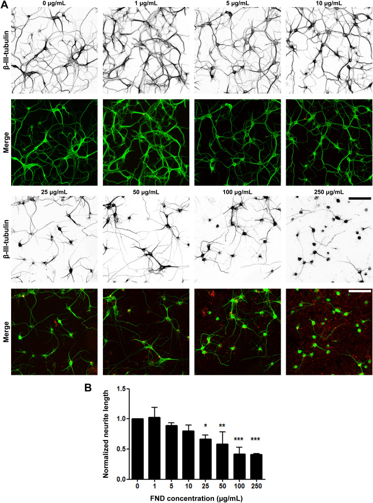
Figure 7. Fluorescent nanodiamonds reduced neurite outgrowth in a dosage-dependent manner in dissociated hippocampal neurons.
(A) Images of dissociated hippocampal neurons treated with various concentrations of FNDs 4 hours after seeding and incubated for 3 days in vitro. Images on the top row show β-III-tubulin staining (inverted to enhance visibility), merged images on the bottom row show FND (red) and β-III-tubulin staining (green). Scale bars represent 100 μm. (B) Quantification of total neurite length per neuron in FND treated hippocampal neurons. All quantification data were obtained from 3 independent repeats and normalized to the group without FND. * p < 0.05, ** p < 0.01, *** p < 0.001 one-way ANOVA followed by Dunnett's post-hoc analysis against the control group (0 μg/mL FND). The bar graph is expressed as mean ± SEM.

