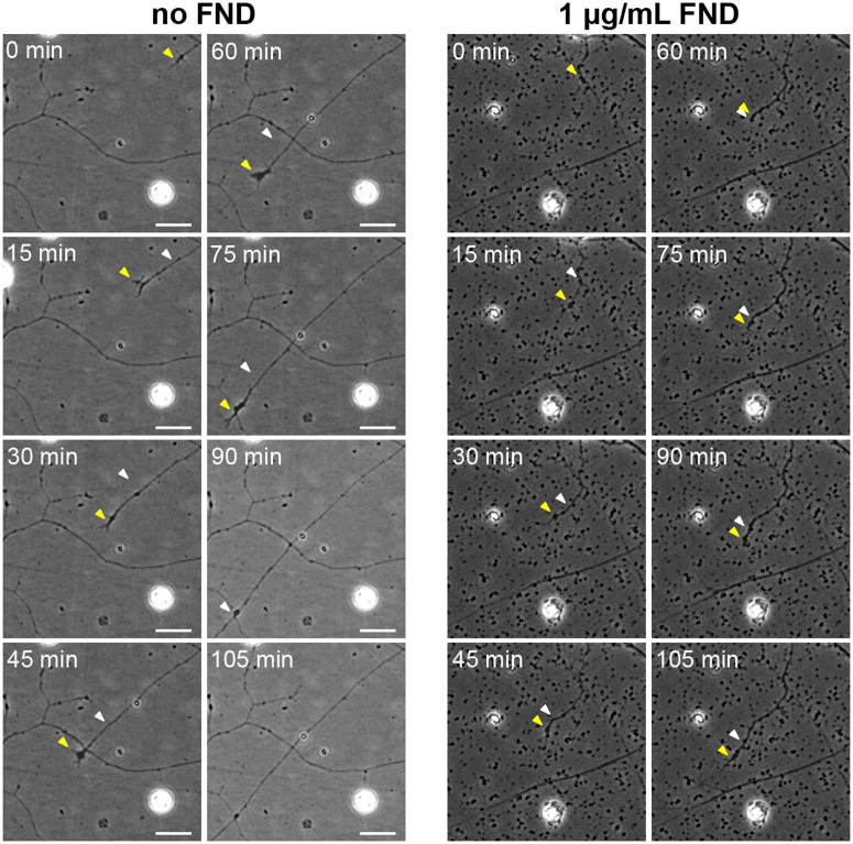
Figure 9. Fluorescent nanodiamond clusters acted as spatial hindrance on advancing neuronal growth cones.
The time-lapse phase contrast image sequences showing the advancing growth cones of DRG neurons under no (left panels) or 1 μg/mL of FND (right panels) treatment for 1 day. The yellow arrowhead points to the growth cone for the current time, and the white arrowhead points to the growth cone at the previous time. All images have the same magnification and all scale bars represent 20 μm.

