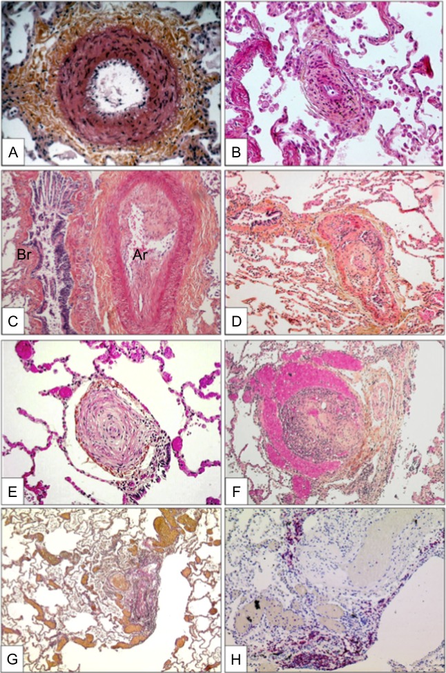
Figure 1.
Characteristic histlogical changes seen in the pulmaonray areriesof lungs affected with PAH showing (A) medial hypertrophy, (B) concentric non-laminar intinal fribrosis, (C) eccentric intimal fibrosis, (D) thrombotic lesions, (E) concentric laminar intimal fibrosis, (F) plexiform lesions of small sinusoid-like vessesls, (G)multiple dilation lesions associated with centrally located plexiform lesions and (H) presence of T-lymphocytes (CD-3 positive) cells in a plexifrom lesion). From Montani el al. 11

