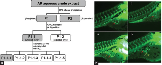
Figure 1.

Schematic diagram showing the method for bioassay-guided fractionation of AR aqueous crude extract and representative images of zebrafish study. (a) Active fractions at each level (shown in gray boxes) were selected for further fractionation. P1-1-1 was the fraction finally selected for mechanistic study. (b) Zebrafish embryos treated with (i) embryo medium only and zebrafish embryos treated with P1-1-1 at (ii) 0.03125 mg/ml, (iii) 0.0625 mg/ml, and (iv) 0.125 mg/ml. Red arrows indicate the smooth basket-like structure of sub-intestinal vessel (SIV) appearing at the bottom of each embryo. Yellow arrows indicate the new blood vessels (sprouts) formed on the SIV of embryos treated with P1-1-1 which were observed in (iii) and (iv)
