
Figure 6. Effect of PLGA-CUR NPs on the AR and β-catenin.
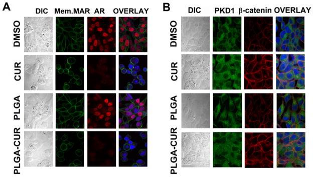
(A) PLGA-CUR NPs lowers the AR expression in C4-2 cells. C4-2 cells were treated with 20 μM curcumin or equivalent PLGA-CUR NPs, or respective control (DMSO or PLGA-NPs) for 24 hrs, immunostained for membrane marker (green), AR (red) and counter-stained with DAPI (blue). Curcumin and PLGA-CUR NPs treated cells showed significantly lower AR staining compared to control C4-2 cells. (B) PLGA-CUR NPs treatment improves the cellular localization of β-catenin and PKD1. C4-2 cells were treated with 20 μM curcumin or equivalent PLGA-CUR NPs, or respective control (DMSO or PLGA-NPs) for 1 hr, immunostained for PKD1 (green), β-catenin (red) and counter-stained with DAPI (blue). CUR and PLGA-CUR NPs treated cells exhibited higher membrane β-catenin staining and localized with PKD1, compared to control C4-2 cells. Original Magnifications 600X.