FIGURE 2.
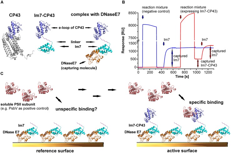
Experimental approach for mapping the binding sites of extrinsic subunits and assembly factors of PSII. The structural models are based on the crystal structures of PSII (Umena et al., 2011; pdb code: 3ARC) and the complex between DNase E7 and Im7 (Ko et al., 1999; pdb code: 7CEI). (A) Structural models for CP43, the e-loop of CP43 tagged with Im7 and the Im7-CP43 fusion protein bound to DNase E7. The Im7-tag (cyan) and DNase E7 (orange) are located in a position occupied by the transmembrane helices of native CP43 (gray), and thus neither the tag nor DNase E7 interfere with the binding of soluble interaction partners to the e-loop (light blue). (B) Preparation of active (red) and reference surface (blue) for surface plasmon resonance (SPR) interaction analysis. Unspecific binding of the reaction mixture to the surface is checked by injecting a 1000-fold dilution over the reference cell, upon which the signal returned to the baseline level. This indicates the absence of unspecific binding. In contrast, injection of a reaction mixture (identical dilution) expressing Im7-CP43 results in stable immobilization of 510 RU of fusion protein. Accordingly, the purity of the immobilized PSII domain on the SPR surface is close to 100%. Finally, both surfaces were saturated with purified Im7 (200 nM) to achieve maximal comparability between reference and active surface; division of the y-axis (high bulk signal caused by the high ionic strength of the immobilization buffer) should be considered. (C) Schematic structures of the surfaces prepared in (B), which were used as positive control for SPR interaction analysis between Im7-CP43 [colors according to (B)] and PsbV (salmon). DNase E7 is covalently bound to the sensor surface, allowing stable immobilization of Im7 and Im7-tagged proteins. Unspecific binding is checked by injection of PsbV on a reference surface with immobilized Im7, whereas the sum of specific and unspecific binding is monitored on an active surface containing the Im7-tagged domain in the required amount. The reference-subtracted binding responses for interaction analysis between PSII domains and PsbV, PsbO or CyanoP are shown in Figures 3–5.

