Figure 1.

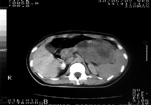
Magnetic resonance imaging shows that the tumor is a well-marginated, large, encapsulated, solid and cystic mass with areas of hemorrhagic degeneration, as revealed by high signal intensity.

Magnetic resonance imaging shows that the tumor is a well-marginated, large, encapsulated, solid and cystic mass with areas of hemorrhagic degeneration, as revealed by high signal intensity.