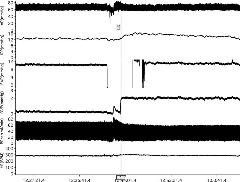
Figure 2.
EVP protocol. Baseline measurements were made in the supine position for ≈10 min. The EVP pipette was then withdrawn from the episcleral vein to prevent breaking the tip while the animal was tilted, then the pipette re-inserted in the vein and measurements continued for ≈10 min in the head-down tilt position.

