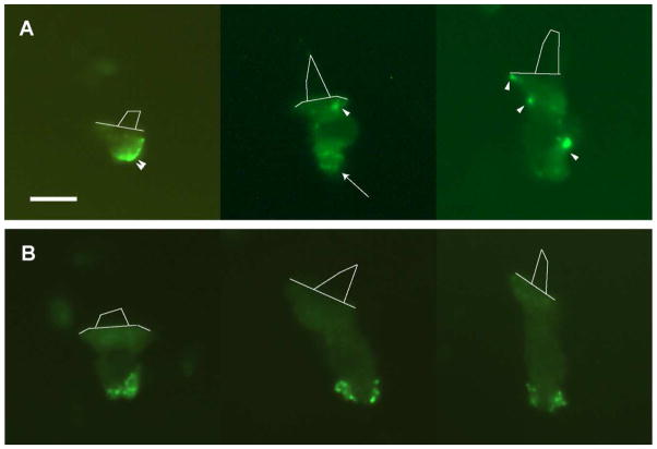
Figure 1. CTX labeling of isolated hair cells reveals punctate GM1-rich microdomains.
Unfixed (A) and pre-fixed (B), dissociated hair cells from throughout the abneural and neural regions of the cochlea exhibited bright, punctate spots following CTX application. Unlabeled preparations and those treated with unconjugated CTX were devoid of punctate staining (not shown). Live cells displayed punctate spots between 0.3 and 1 μm in diameter (arrow) as well as large, diffuse domains along the basolateral surface (arrowhead). Punctate spots were exclusively found on pre-fixed, CTX-labeled cells. Hair bundles are outlined with thin lines to show cell orientation. Scale bar = 10 μm.

