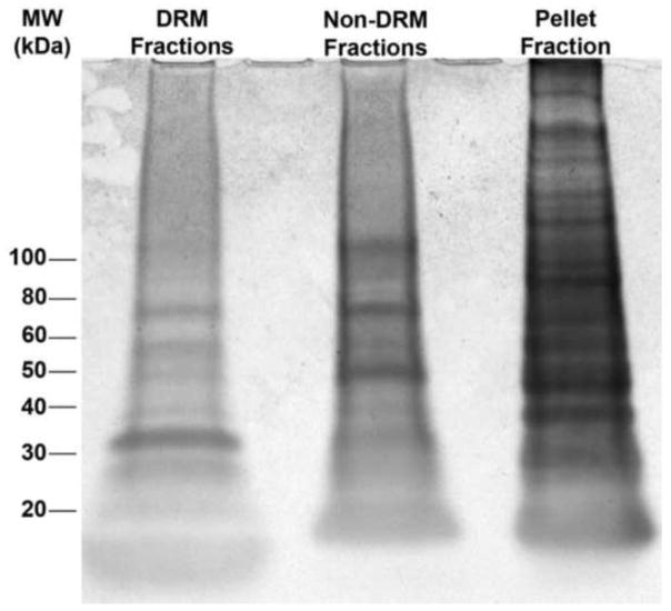
Figure 6. SDS-PAGE of cochlear fractions reveals heterogeneous protein distributions among DRM, non-DRM, and soluble protein pools.
Whole cochlea lysates treated with 1% TX-100 were separated by SDS-PAGE and stained using Coomassie Blue to reveal the diversity and abundance of proteins in pooled DRM fractions (4–6), pooled detergent sensitive fractions (9–11), and the pellet of soluble proteins.

