
Figure 7.
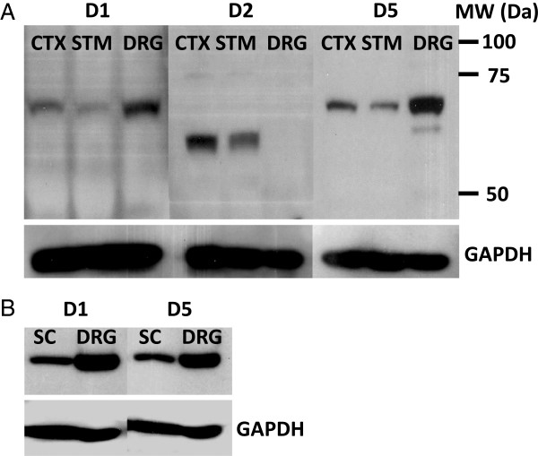
Western blotting of dopamine receptors. Immunoblots for D1, D2, and D5 dopamine receptors were obtained for frontal cortex (CTX), striatum (STM), spinal cord (SC) and dorsal root ganglia (DRG) proteins. All wells were loaded with the same concentration of total protein with GAPDH serving as an internal control. A) Membranes were probed with antibodies specific for D1, D2, or D5 dopamine receptors. D1 and D5 were identified in CTX, STM, and DRG as single bands with apparent MW’s of 68 kDa. D2 dopamine receptor protein was identified in CTX and STM as a single band with an apparent MW of 62 kDa. Corresponding sections from each blot were probed with antibody against GAPDH (shown beneath the membranes in A). B) Western blotting of D1 and D5 dopamine receptors were repeated comparing equal concentrations of SC and DRG protein. Corresponding sections from each blot were probed with antibody against GAPDH (shown beneath the membranes in B).