
Figure 9.
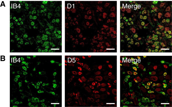
Expression of dopamine receptors in intact dorsal root ganglia. Frozen sections, 20 μm thick, were prepared from lumbar DRG taken from P14 to P28 Sprague Dawley rats perfused with formaldehyde. Sections of dorsal root ganglia were stained with FITC-labeled lectin IB4 (green channel) and monoclonal antibodies to either D1 (red channel, A) or D5 dopamine receptors antibodies (red channel, B), and secondary antibodies against mouse IgG labeled with Alexa 594. Merging the images indicated some co-localization of IB4 and either D1 or D5 dopamine receptors (yellow). Scale bars are 50 μm.