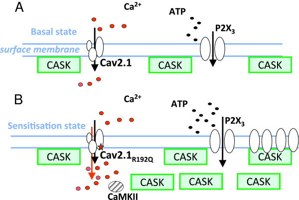
Figure 3.

Proposed mechanism of action of CASK/P2X3 complex in trigeminal sensory neurons. Schematic representation of CASK, P2X3, and CaV2.1 channel protein interactions at the plasma membrane of trigeminal sensory neurons. A, In WT ganglion cells, only a fraction of P2X3 receptors is thought to be associated with CASK that ensures their stable expression. Under these conditions, CaV2.1 channels provide only a minor contribution to Ca2+ influx, and the dynamic CASK/P2X3 association ensures physiological responses and P2X3 receptors turn-over [6]. B, In KI ganglia, the hyperfunctional CaV2.1 channels result in a stronger Ca2+ influx, larger CaMKII activity, and more abundant CASK/P2X3 receptor complexes with improved stability of P2X3 receptors. Sub-membrane vesicle-associated CASK, potentially associated with P2X3 receptor trafficking, is also indicated. Larger release of extracellular ATP [20] facilitates P2X3 responses and may contribute to the process of sensitization of P2X3 receptors.
