Figure 1.
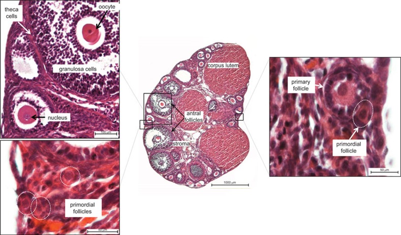
Histological features of the normal post-pubertal ovary. These images show the structures of a post-pubertal mouse ovary; salient features of the human ovary are essentially the same. Major structures include follicles, which house the oocytes, and the post-ovulatory correlate of the follicle, the corpus luteum. Multiple follicles at various stages of development are present within the pre-senescent ovary at any given time. Several stages of follicles can be seen in this section, including the developmentally earliest stages, primordial and primary follicles (lower left and right insets), and the more advanced antral follicles (center and upper left inset). Oocytes are encased in follicular steroidogenic cells, the inner granulosa and the outer theca cell layers (upper left inset); theca interna and theca externa are not clearly depicted in this image but can be seen in Figure 4. Mature follicles and corpora lutea produce estrogen and progesterone, respectively, which are required sequentially for uterine preparation and maintenance of pregnancy. The sample is stained with hematoxylin and eosin. Scale bars: 100 µm (upper left); 50 µm (lower left); 1000 µm (center); 50 µm (right).

