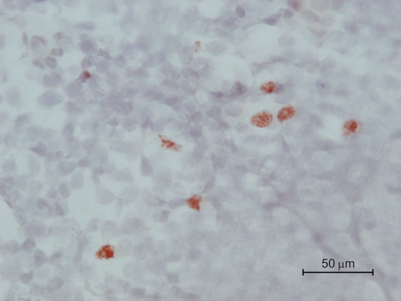
Figure 2.
AIRE expression in mTEC of mouse thymic medulla. Immunohistochemical analysis of thymic medulla of a Balb/c mouse using a monoclonal anti-mouse Aire antibody. mTEC, although not universally Aire+, are distinguished by high cytoplasm/nucleus ratio; thymocytes are smaller cells with low cytoplasm/nucleus ratio. Red/brown stain indicates nuclear staining of Aire protein. Counterstain, hematoxylin. Scale bar=50 µm. AIRE, autoimmune regulator; mTEC, medullary thymic epithelial cell.

