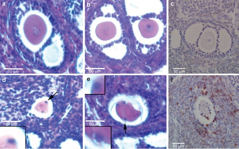
Figure 3.
T lymphocyte infiltration into ovarian follicles of Aire-deficient mice. (a–c) Follicles from WT mice; (d–f) Follicles from Aire-deficient mice. (a, b, d, e) Hematoxylin and eosin stain; (c, f) Immunohistochemistry using anti-CD3 pan-T-cell antibody and hematoxylin counterstain. WT follicles lack detectable lymphocytes, while follicles from Aire-deficient mice are surrounded by T cells, including those in close contact with the degenerating oocyte. Arrowheads in d and e identify putative lymphocytes surrounding oocyte; CD3-positive cells can similarly be seen in proximity to the egg in f. AIRE, autoimmune regulator; WT, wild-type.

