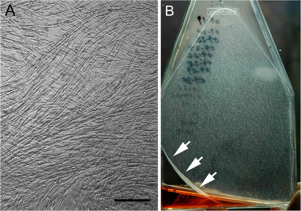
Figure 1.

Representative hyperconfluent cell sheets: Phase contrast microscopy of 4th passage hyperconfluent cell sheet (“HCS”; A), 10X objective magnification, bar = 100 μm. Gross appearance of a representative hyperconfluent 4th passage cells sheet at commencement of spontaneous sheet contraction (arrows), indicating time for sheet harvest to synthesize tensioned synoviocyte bioscaffolds (B).
