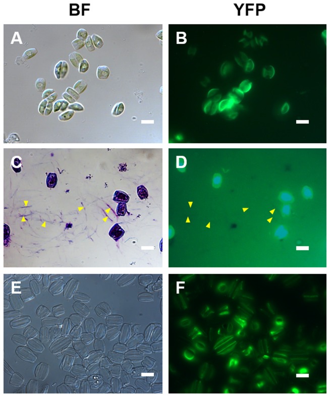
Figure 3. Localization of AC3362-YFP in A. coffeaeformis.
(A, C, E) Bright field microscopy images, and (B, D, F) corresponding epifluorescence microscopy images. (A, B) Live cells. (C, D) Cells after treatment with the polycationic dye ‘Stains-All’. For orientation some trails are labeled by arrowheads. Image (D) was deliberately overexposed to check for YFP fluorescence in the trails. (E, F) Isolated cell walls. Bars = 10 µm.

