
Figure 5. Biosilica association of AC3362-YFP.
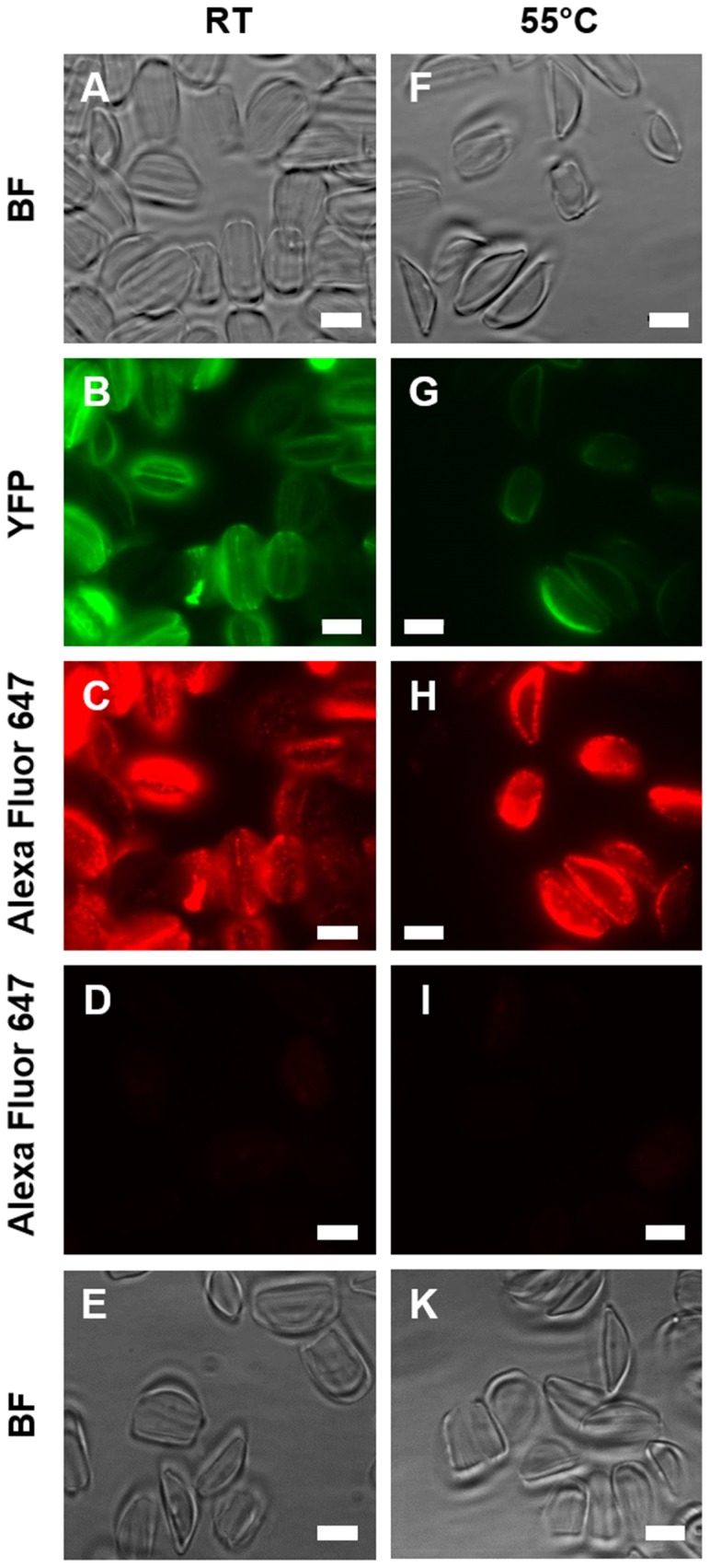
Isolated cell walls (RT) and isolated cell walls after hot SDS teatment (55°C) were analyzed by bright field microscopy (BF), by direct fluorescence microscopy (YFP), and by indirect immunofluorescence microscopy (Alexa Fluor 674) using an anti-YFP primary antibody and an Alexa Fluor 647-labeled secondary antibody. (A-C) Cell walls from a transformant clone expressing AC3362-YFP, and (F-H) cell walls from the same clone after extraction with a hot SDS solution. Cell walls from wild type (D, E) before and (I, K) after extraction with a hot SDS solution. Bars = 5 µm.