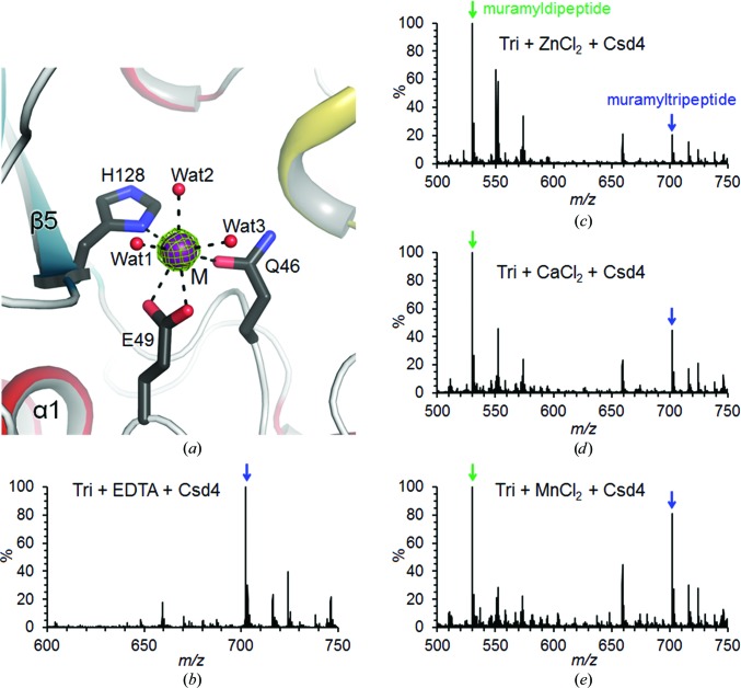
Figure 2.
Metal-dependency of H. pylori Csd4 as a carboxypeptidase. (a) Ribbon diagram of the active site in the Csd4-unbound structure coloured as in Fig. 1 ▶(a). The OMIT mF o − DF c map (contoured at 5σ) for the calcium ion is coloured green. (b–e) Mass spectra of muramylpeptides after the reaction catalyzed by Csd4 in the presence of EDTA (b) or in the presence of Zn2+ (c), Ca2+ (d) or Mn2+ (e). Green and blue arrows indicate the peaks corresponding to the muramyldipeptide and muramyltripeptide (Tri), respectively.

