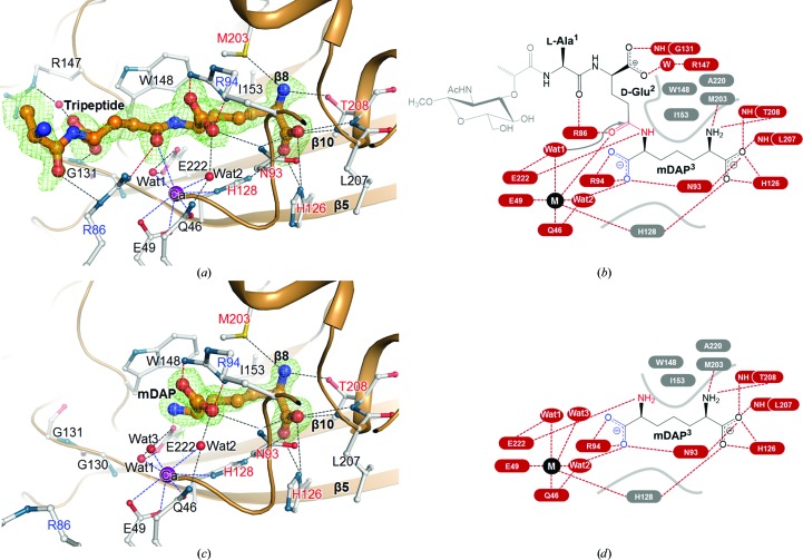
Figure 4.
Ligand interactions in the Csd4–muramyltripeptide and Csd4–mDAP structures. (a, c) Ribbon diagrams of the active site in the Csd4–muramyltripeptide (a) and Csd4–mDAP (c) structures. The tripeptide and mDAP are shown as stick models. Since the sugar moiety of the NAM-tripeptide was invisible, only the tripeptide portion has been modelled into the electron-density map. Residues interacting with the tripeptide or mDAP are shown as stick models. The OMIT mF o − DF c maps for the tripeptide (contoured at 1.5σ) and mDAP (contoured at 2.5σ) are coloured green. The red and blue dotted lines indicate the interactions of the tripeptide or mDAP with the metal ion and two key Arg residues (Arg86 and Arg94), respectively. (b, d) Schematic diagrams of interactions of the bound muramyltripeptide (b) and mDAP (d) with Csd4. The red and grey labels represent electrostatic and hydrophobic residues interacting with these ligands, respectively.

