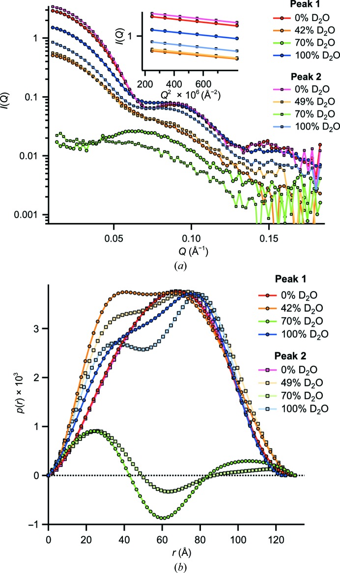
Figure 4.
SANS curves and p(r) functions of peak 1 and peak 2. (a) SANS curves at four different contrast conditions at 20°C. All data sets are drawn without applying scaling. The total protein concentrations of all samples were identical (4.5 mg ml−1). Guinier fits to the 0, 42 (49) and 100% D2O data are shown as an inset (the 70% D2O data were not fitted using the Guinier approach owing to their elevated noise level). (b) Pair-distance distribution functions p(r) of the SANS data in arbitrary units generated with GNOM (Svergun, 1992 ▶). The two 0% D2O data sets are very similar (cf. Supplementary Fig. S6). The 0, 42, 49 and 100% D2O data are normalized to the second peak and the 70% D2O data to the first peak (using a different scaling factor for clarity).

