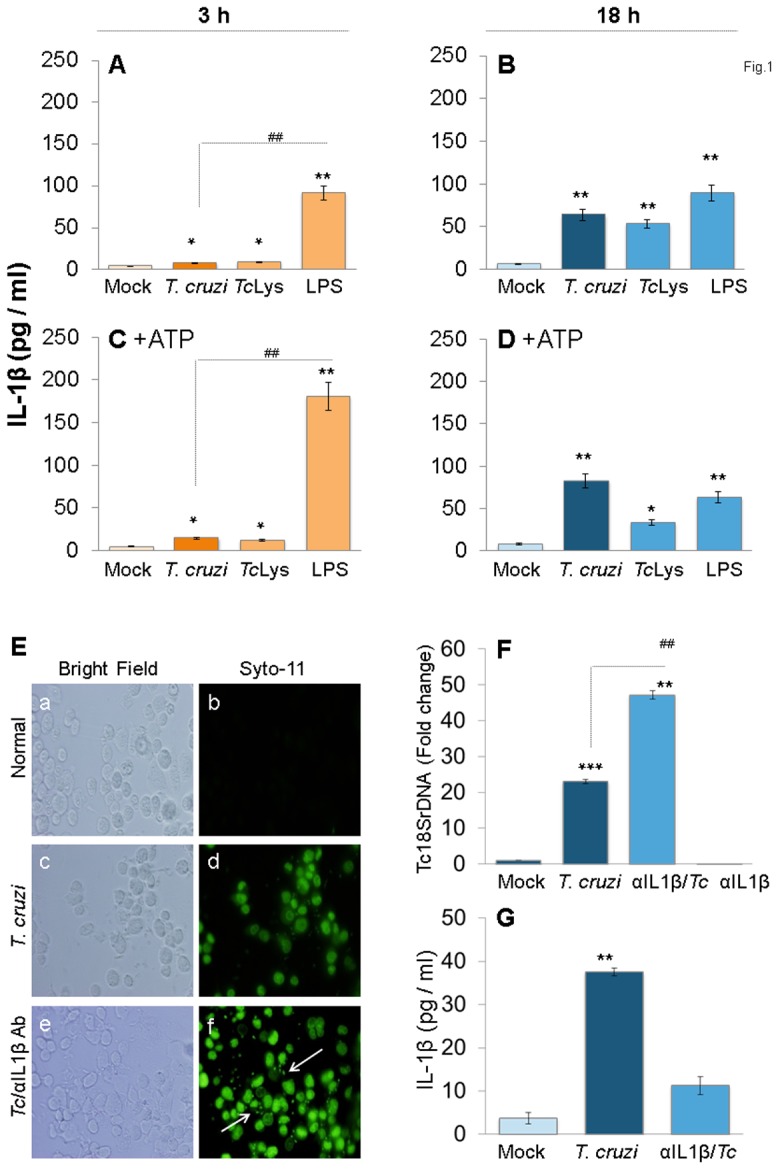
Figure 1. IL-1β production in macrophages infected by T. cruzi.

(A–D) PMA-differentiated THP-1 mφs were incubated with T. cruzi trypomastigotes (cell: parasite ratio, 1∶3), Tc lysate (10 µg protein/106 cells) or LPS (100 ng/ml) for 3 h (A&C) and 18 h (B&D). In some experiments, ATP was added during last 30 min of incubation (C&D). IL-1β release in supernatants was determined by ELISA. (E–G) IL-1β contributes to parasite control in mφs. THP-1 mφs were incubated with SYTO®11-labeled T. cruzi in the presence or absence of anti-IL-1β antibody for 18 h. (E) SYTO®11 fluorescence as an indicator of parasite uptake (shown by arrows) was determined by using an Olympus BX-15 microscope equipped with a digital camera (magnification 40X). (F) Quantitative PCR analysis of parasite burden in infected mφs by using Tc18SrDNA-specific oligonucleotides (normalized to human GAPDH). (G) Addition of anti-IL-1β antibody depletes secreted IL-1β levels in T. cruzi-infected mφs. In all figures, data are representative of three independent experiments and presented as mean ± SD. Significance is shown by *normal versus infected and #treated/infected versus infected (*,#p<0.05, **,##p<0.01, and ***,###p<0.001).
