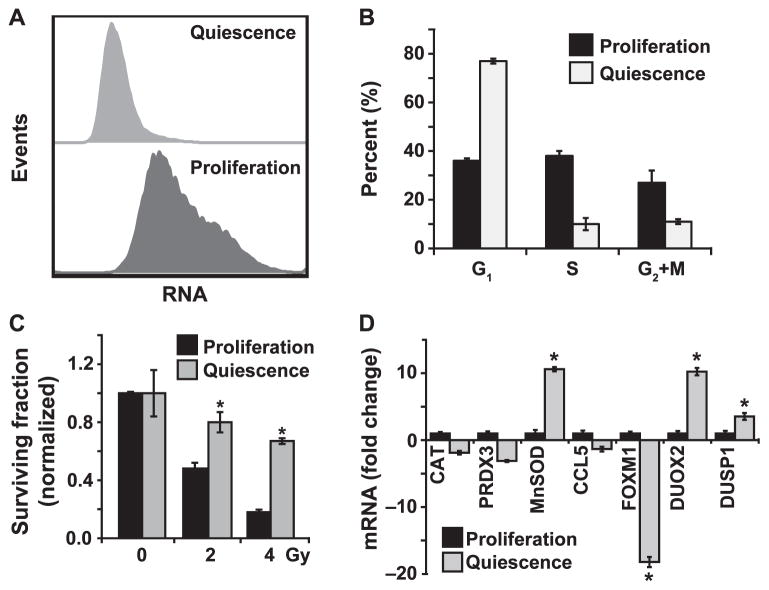
FIG. 1.
Quiescent HNSCC are radioresistant and have lower basal FOXM1 expression. Panel A: Flow cytometry assay to assess quiescence. Ethanol-fixed Cal27 cells representative of low and high proliferative index were incubated with Hoechst 33258 (for DNA) and Pyronin Y (for RNA), and fluorescence analyzed by Aria II flow cytometer. Panel B: Flow cytometry measurements of DNA content to examine cell cycle phase distributions. Cultures with a lower proliferative index (S phase <10%; G1 phase >75%) are considered as quiescent and cultures with a higher proliferative index (S phase >30%; G1 phase <40%) are considered as proliferating cells. Panel C: A clonogenic assay designed to accommodate post-lethal damage repair was used to examine surviving fraction in Cal27 cells. Panel D: A Q-RT-PCR assay was used to measure mRNA levels in proliferating and quiescent Cal27 cells. Fold change in mRNA levels in quiescent cells was calculated relative to corresponding mRNA levels in proliferating cells. Asterisks represent statistical significance of quiescent compared to proliferating cells. n = 3; P < 0.05.

