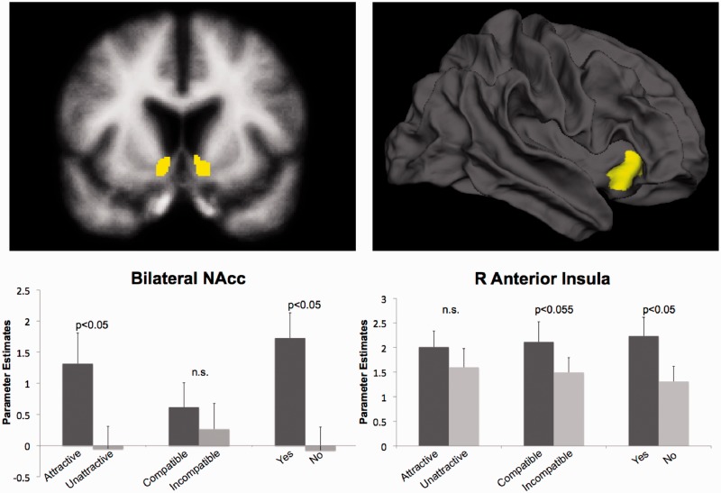
Fig. 2.
(Bottom) Activity of the a priori ROIs from Interval 2. (Top) Brain images showing the anatomically defined ROIs created with FreeSurfer’s parcellation algorithm. The Yes/No and Compatibility/Attractiveness parameter estimates are drawn from separate FIR models (see Methods: fMRI Data Analysis for details). Error bars represent standard error of the mean.

