Figure 1.
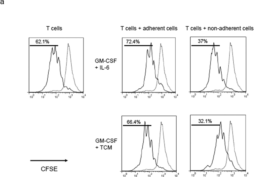
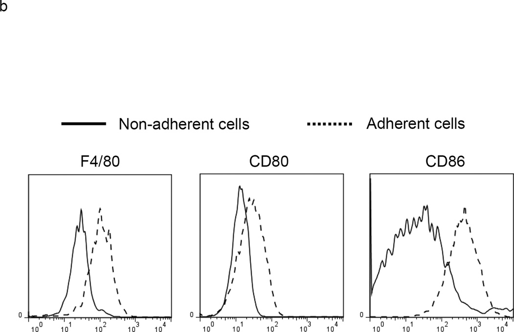
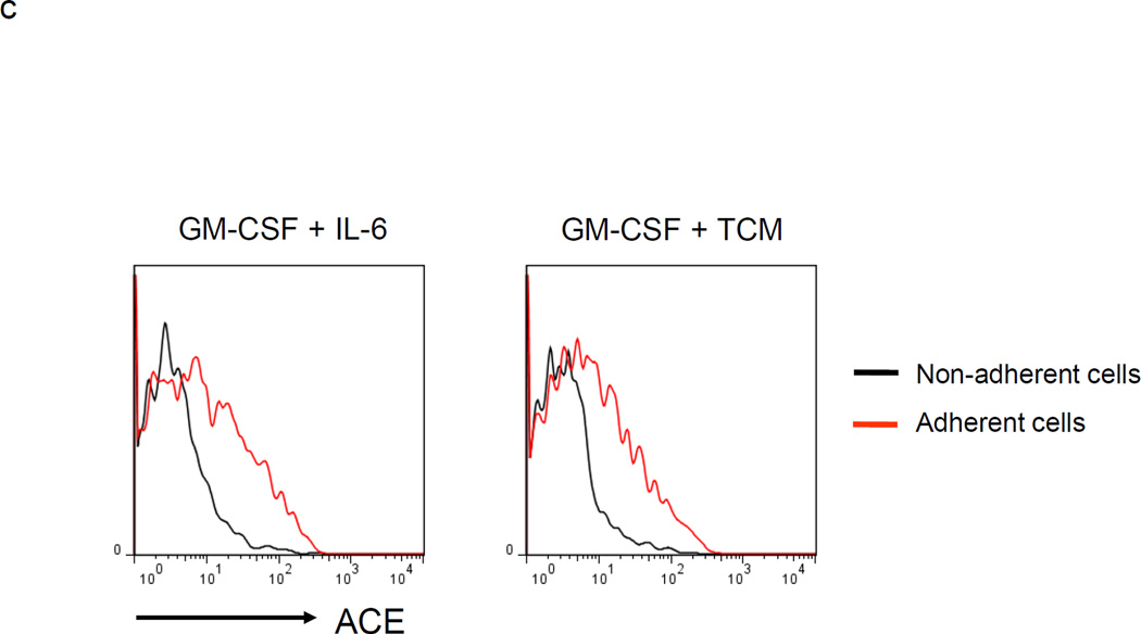
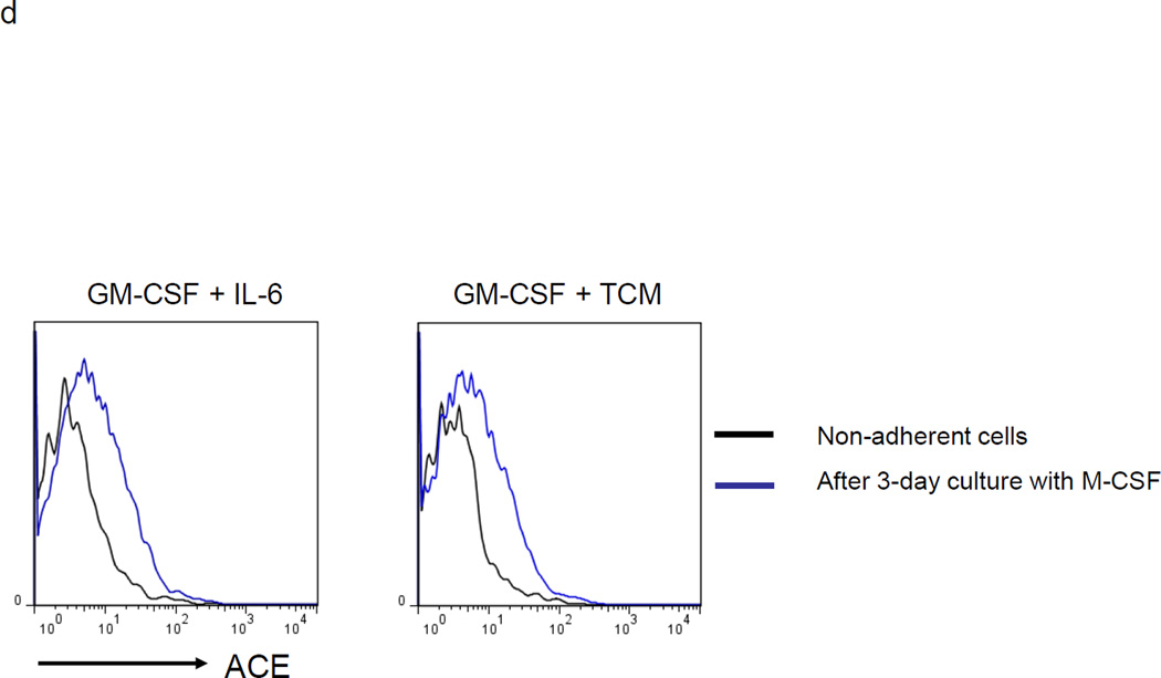
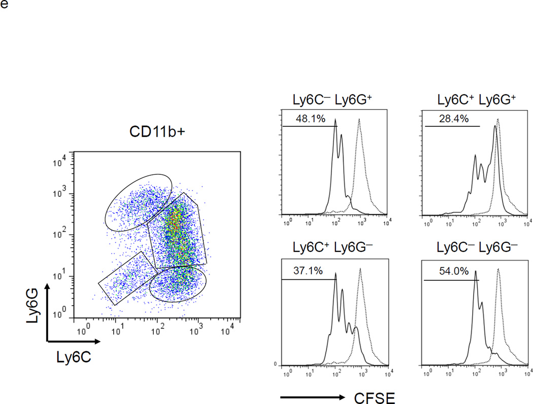
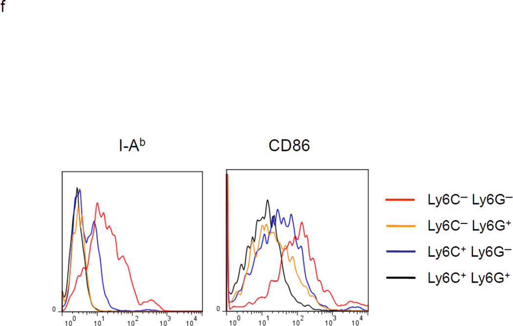
MDSC development in vitro. a. WT BM cells were cultured with GM-CSF and either IL-6 or tumor-conditioned medium (TCM) for 4 days. The non-adherent and adherent cells were then harvested and separately co-cultured with anti-CD3 and anti-CD28 stimulated CFSE-labeled T cells for another 3 days. Typical T cell proliferation profiles are shown (line) compared to the original T cells (dotted). The non-adherent cells suppress T cell proliferation resulting in less dilution of CFSE. b. The expression of surface maturation markers F4/80, CD80 and CD86 was assessed on the non-adherent and adherent cells. c. ACE expression on the non-adherent and adherent cells. d. ACE expression on the non-adherent cells with and without culture in M-CSF. e. The surface expression of Ly6G and Ly6C was measured on non-adherent cells (left). Four populations were identified: Ly6C−Ly6G+, Ly6C+Ly6G+, Ly6C+Ly6G−, and Ly6C−Ly6G−. These populations were gated and sorted, and their immunosuppressive activities were assessed by their ability to inhibit T cell proliferation (right). f. The surface maturation markers MHC class II (I-Ab) and CD86 were evaluated on the gated cells. For all the experiments, the representative flow cytometry figures presented are representative of at least 3 independent experiments.

