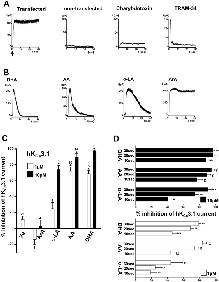
Figure 2. Membrane expression of cloned human KCa3.1 in HEK-293 in inside-out patches and basic pharmacological characterization.
A) From left to right: Exemplary traces of immediate activation of hKCa3.1-outward currents upon excision of the patch into 3 µM Ca2+-containing bath solution (as indicated by arrow). KCa-outward currents are absent in non-transfected HEK-293. Inhibition of hKCa3.1-outward currents by charybdotoxin (100 nM, in the pipette solution) and TRAM-34 (1 µM, in the bath solution). B) Inhibition of hKCa3.1 by ω3 and arachidonic acid. From left to right: Time course of inactivation of hKCa3.1 by docosahexaenoic acid (DHA, 10 µM), arachidonic acid (AA, 10 µM), α-linolenic acid (α-LA, µM) over time. Saturated arachidic acid (ArA, 10 µM) did not affect channel activity. C) Concentration-dependence of inhibition. Note that half of the current was inhibited by AA, DHA, and α-LA at approx. 1 µM. D) Time course of channel inactivation by two concentrations of AA, DHA, and α-LA over time. Data are means ± SEM (% inhibition of KCa3.1-current normalized to initial peak amplitude after patch-excision); numbers in the graphs indicate the number of inside-out experiments; *P<0.05 vs. vehicle (Ve); One-way ANOVA and Tukey post hoc test.

