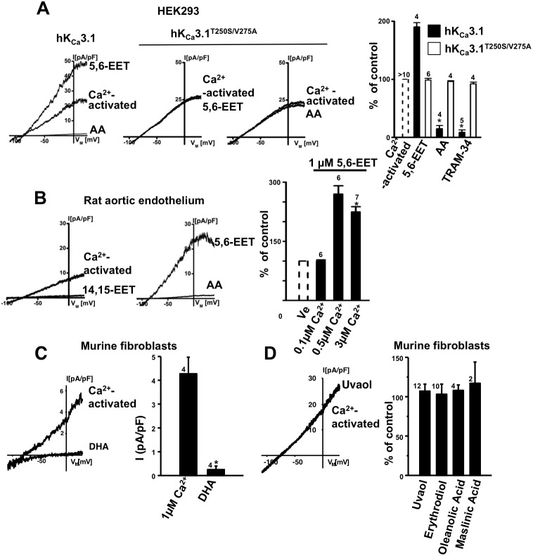
Figure 6. 5,6-EET-potentiation of KCa3.1 currents.
A) Whole-cell current traces; from left to right: potentiation of Ca2+-pre-activated hKCa3.1 by 5,6-EET (1 µM) followed by inhibition of the current by AA (10 µM), insensitivity of the hKCa3.1T250S/V275A mutant to 5,6-EET, and insensitivity of the hKCa3.1T250S/V275A mutant to AA (10 µM) and TRAM-34 (1 µM). The hKCa3.1 currents were pre-activated by 250 nM Ca2+. Panel on the right: summary data. B) From left to right: Ca2+-pre-activation of rat endothelial rKCa3.1 by 3 µM Ca2+ and current inhibition by 14,15-EET (1 µM), larger currents in the presence of 5,6-EET (1 µM) and inhibition by AA (10 µM). Panel on right: Summary data: dependence of 5,6-EET-potentiation on the intracellular Ca2+. Note that at a low intracellular Ca2+ (0.1 µM) that is below/near the threshold for KCa3.1 activation, 5,6-EET did not potentiate the current. In contrast, potentiation occurred at an intracellular Ca2+ concentration that is near the EC50 for Ca2+-activation of KCa3.1 as well as at a saturating Ca2+ concentration. C) DHA (1 µM) blocked Ca2+-pre-activated mKCa3.1 in murine fibroblasts. D) Pentacyclic triterpenes did not modulate murine fibroblast mKCa3.1 at a concentration of 1 µM. Data are means ± SEM (% inhibition of KCa3.1-current normalized to initial peak amplitude after establishing electrical access (by seal rupture) and stable Ca2+-activation of KCa3.1-outward currents); Numbers in the graphs indicate the number of whole-cell experiments; *P<0.05 vs. control (peak amplitude of the KCa3.1-current in the respective cell); One-way ANOVA and Tukey post hoc test.

