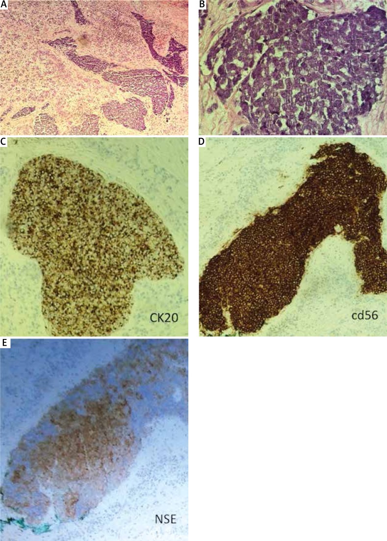
Figure 2.
Merkel cell carcinoma. A, B – Hematoxylin and eosin staining. The obtained result of the histopathological examination found within the dermis area is low-differentiated small cancer cells with scanty cytoplasm and round nuclei with small grains. Necrosis was seen and a number of apoptotic cells and the figure division. In the stroma around tumors, there were seen numerous blood vessels and infiltration of inflammatory cells. C – Positive cytokeratin 20 staining. D – Positive CD56 staining. E – Positive neuron-specific enolase (NSE) staining

