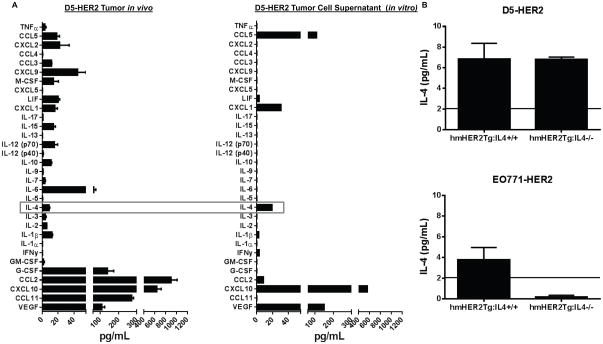
Figure 1. Characterization of cytokines/chemokines expressed by D5-HER2 in vitro and in vivo.
A. Expression of 32 cytokines/chemokines in the microenvironment of D5-HER2 tumors was analyzed using a luminex-based approach. n=5 animals. B. A representative characterization of the cytokine/chemokine expression profile of D5-HER2 cells grown in vitro. Cell supernatant was harvested after 72 hours of growth and analyzed using a luminex-based approach. C. Evaluation of IL4 production by tumors grown in hmHER2Tg:IL4+/+ or hmHER2Tg:IL4−/− animals. 3×10³ D5-HER2 or 1×10⁶ EO771-HER2 cells were inoculated subcutaneously in the flank of indicated animals. Tumors were harvested, homogenized and IL4 was quantified using ELISA. Solid line represents the limit of detection of the assay. n=5 animals per group. Error bars represent standard error of the mean (SEM).

