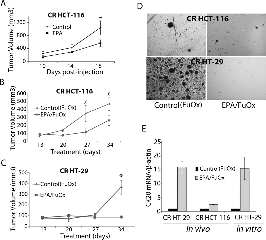
Figure 4.
Inhibition of CR HCT-116 (A&B) or CR HT-29 cells (B) xenografts in EPA and/or FuOx treated SCID mice. A: EPA was administered for 7 days before inoculation of 1×10⁶ CR HCT-116 cells and continued for the duration of the experiment. B&C: EPA+FuOx treatments were started 7 days after CR cells inoculation. Each data point represents average of 8 tumors ± SE. * p >0.05. D: Colonosphere formation in the cells isolated from CR HCT-116 and CR HT-29 xenografts ×100. E: qPCR on RNA from tumors of CR HT-29 and CR HCT-116 cells and CR HT-29 cells.

