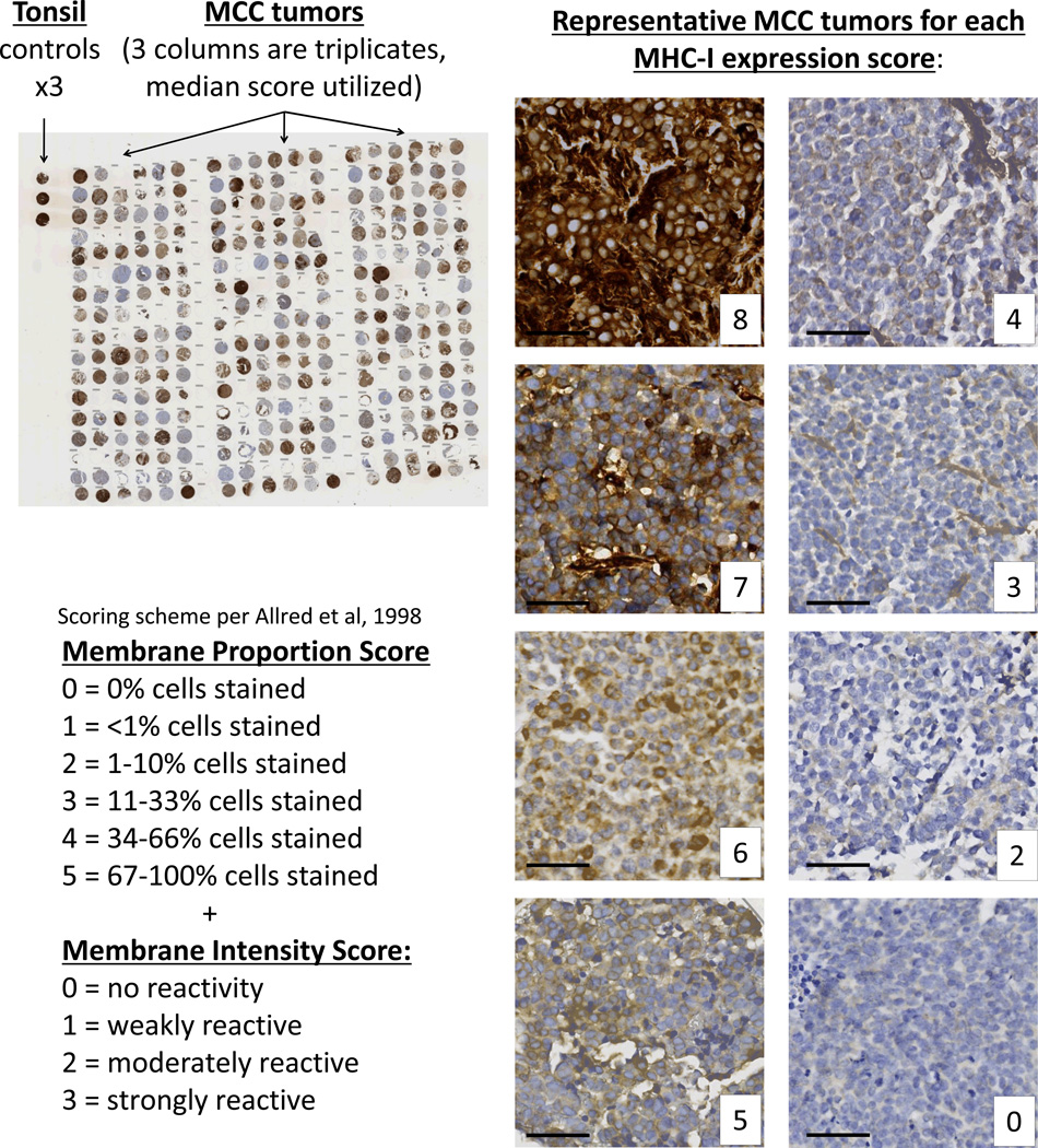
Figure 3. MHC class I immunohistochemistry of Merkel cell carcinoma tumors.
A total of 114 MCC tumors were represented on 5 tissue microarrays and were stained with the EMR8-5 antibody that recognizes HLA-A, -B, and -C. These were scored for proportion of cells expressing MHC class I and intensity of the expression utilizing the Allred scoring system ranging from 0 to a maximal score of 8. Representative MCC tumors at each combined Allred score are shown at right. Black bar represents 50 micrometers.

