Figure 2.
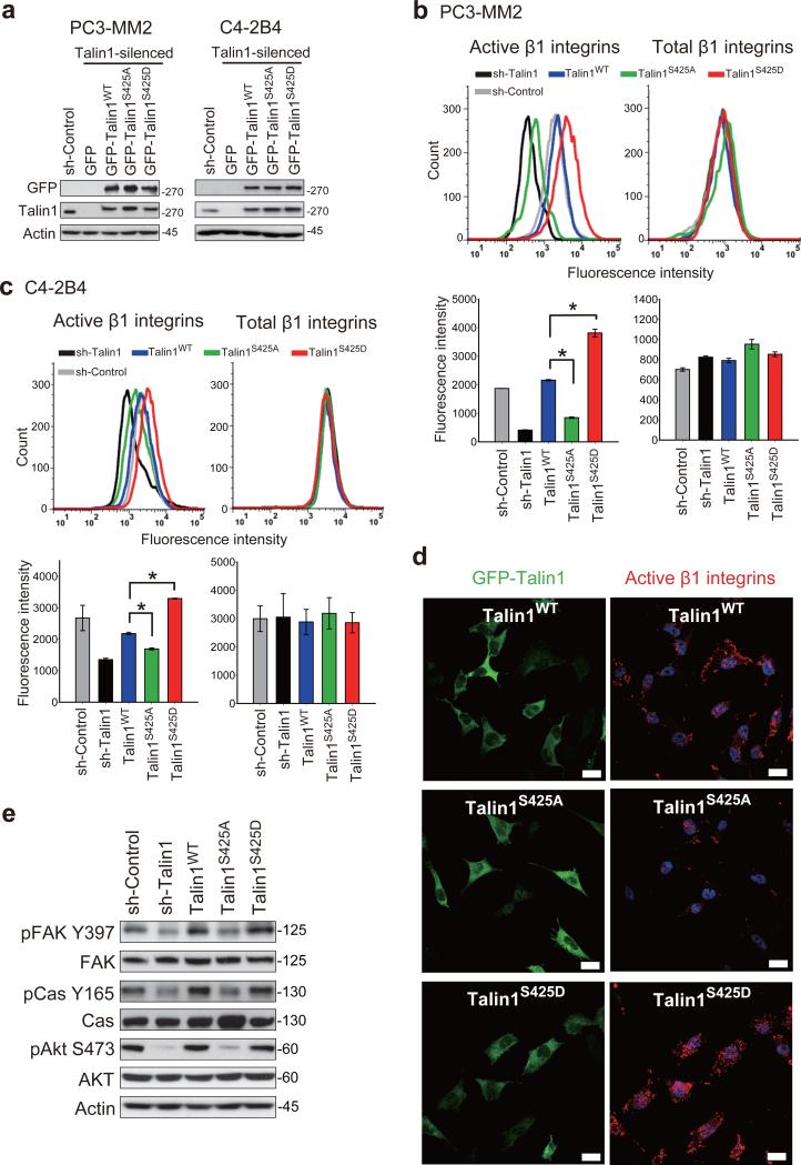
Talin1 S425 phosphorylation promotes β1 integrin activation. (a) Expression of GFP (empty vector), GFP-talin1WT and specified GFP-tagged talin1 mutants in talin1-silenced PC3-MM2 and C4-2B4 cells. (b-c) Flow cytometric analysis of activated and total β1 integrins in (b) PC3-MM2 and (c) C4-2B4 cells expressing talin1WT and mutants (top panels). Quantitation of fluorescence intensity is shown in the bottom panels. *P < 0.005. (d) Immunofluorescence staining of GFP and activated β1 integrins in PC3-MM2 cells expressing talin1WT and mutants. Nuclei were counterstained by Hoechst 33342. Scale bar represents 25 μm. (e) Immunoblotting of pFAK Y397, total FAK, pp130 Cas Y165, total p130 Cas, pAkt S473 and total Akt in PC3-MM2 cells expressing talin1WT and mutants.

