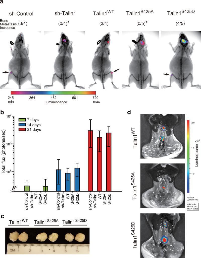
Figure 5.
Talin1 S425 phosphorylation promotes bone colonization in vivo. (a) Luciferase-labeled PC3-MM2 sh-control, talin1-silenced, talin1WT and mutant-expressing cells (1 × 10⁶) were injected into nude mice intracardially. After 21 days injection, bioluminescence and X-ray imaging of mice were superimposed to localize tumor growth. Arrow indicates tumor growth in femur/tibia junction. *Compared to talin1WT, P < 0.05. (b) Tumor burden in the femur/tibia of mice following intracardiac injection. Tumor growth was estimated by luciferase activity (presented as photons/sec). (c-d) PC3-MM2 talin1WT and mutant-expressing cells (5 × 10⁵) were orthotopically injected into the prostate. (c) Sample tumor sizes of lymph node metastases. (d) Bioluminescence imaging of lymph node metastases of PC3-MM2 talin1WT and mutant-expressing cells in mice after removing the primary tumor.

