
FIGURE 2.
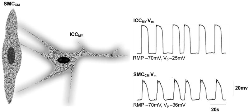
Schematic diagram of an Interstitial Cell of Cajal (ICC) and an associated smooth muscle cell (SMC) of the circular muscle (CM) along with membrane potential (Vm) traces from an ICC and SMC. Both the ICC and SMC membrane activities demonstrate a periodicity of 3 cpm. The amplitude of the ICC membrane potential (~45 mV) is higher than that of the SMC (~34 mV). The membrane potential traces are reproduced from Ref 11.