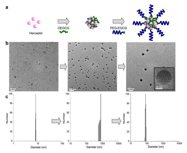
Fig. 1. Schematic diagram and morphology of self-assembled MNC loaded with proteins.
a, Schematic diagram of the self-assembly process to form the MNC. The MNC was formed through two sequential self-assemblies in an aqueous solution: Complexation of OEGCG with proteins to form the core followed by complexation of PEG-EGCG surrounding the pre-formed core to form the shell. b, TEM images and c, hydrodynamic size distributions of complexes observed at each step of self-assembly.

