Figure 1.
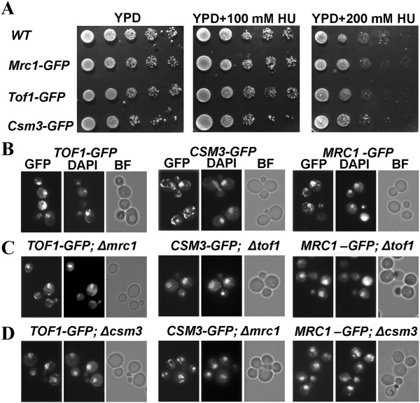
Independent nuclear localization of the subunits of the Tof1/Csm3/Mrc1 complex. (A) Viability test of the S-phase checkpoint GFP-tagged proteins by 10-fold serial dilution assay. 5 μL of each dilution are spotted onto YPD, YPD supplemented with 100 mM and 200 mM HU. (B-D) All GFP strains are paraformaldehyde fixed and subjected to fluorescent microscopy analysis to detect the position of GFP-tagged proteins in the cell. 2.5 μg/ml DAPI staining is used for all of the probes to visualize the position of nucleus. The obtained GFP and DAPI signals are analyzed for co-localizations. GFP - Filter set 38HE (Zeiss); DAPI - Filter set 01 (Zeiss); BF – bright field.

