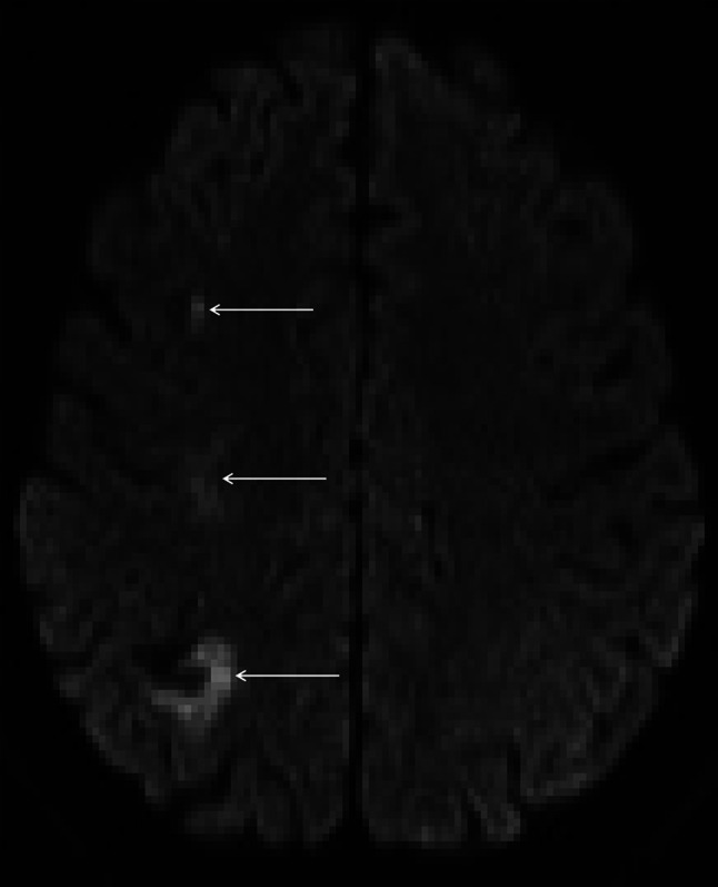
Fig. 3.

Axial DWI reveals multiple areas of infarction axial DWI reveals ischaemic damage in the right superior parietal lobe, right centrum semiovale and middle frontal sulcus (white arrows) suggesting involvement of the internal (subcortical) borderzone territories. In addition there is a small area of gyral haemosiderin staining within the right parietal lobe close to the infarct
