
Figure 4.
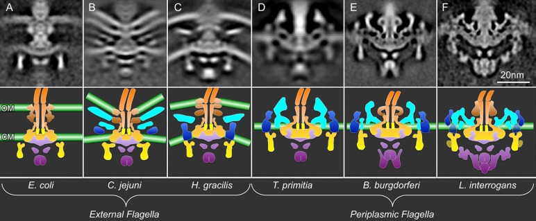
In situ flagellar motor structures from different organisms determined by cryo-ET and subtomogram averaging. (A–C) External flagella: E. coli (EMDB-5311), C. jejuni (EMDB-5300), and H. gracilis (EMDB-5309) flagellar motors. (D–F) Periplasmic flagella: B. burgdorferi (EMDB-5627), T. primitia (EMDB-1235), and L. interrogans (EMDB-5912–5914) flagellar motors.51 The bottom panels show the corresponding cartoon models. The color scheme is the same as that in Figure 3. Noticeably, the densities of putative stator and export apparatus are clearly revealed in some bacterial species.