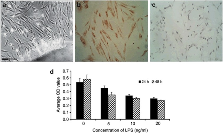
Figure 5.
Characterization of HPDLFs by histological examinations with scanning electron microscopy (a). Immunohistochemistry analysis: vimentin-positive (b) and cytokeratin-negative (c) cells in the overlay were identified as differentiating HPDLFs. The HPDLFs were treated with various doses of LPS for 24 h or for 48 h, and then, cell proliferation was analyzed by MTT assay (d). Scale bar=100 μm.

