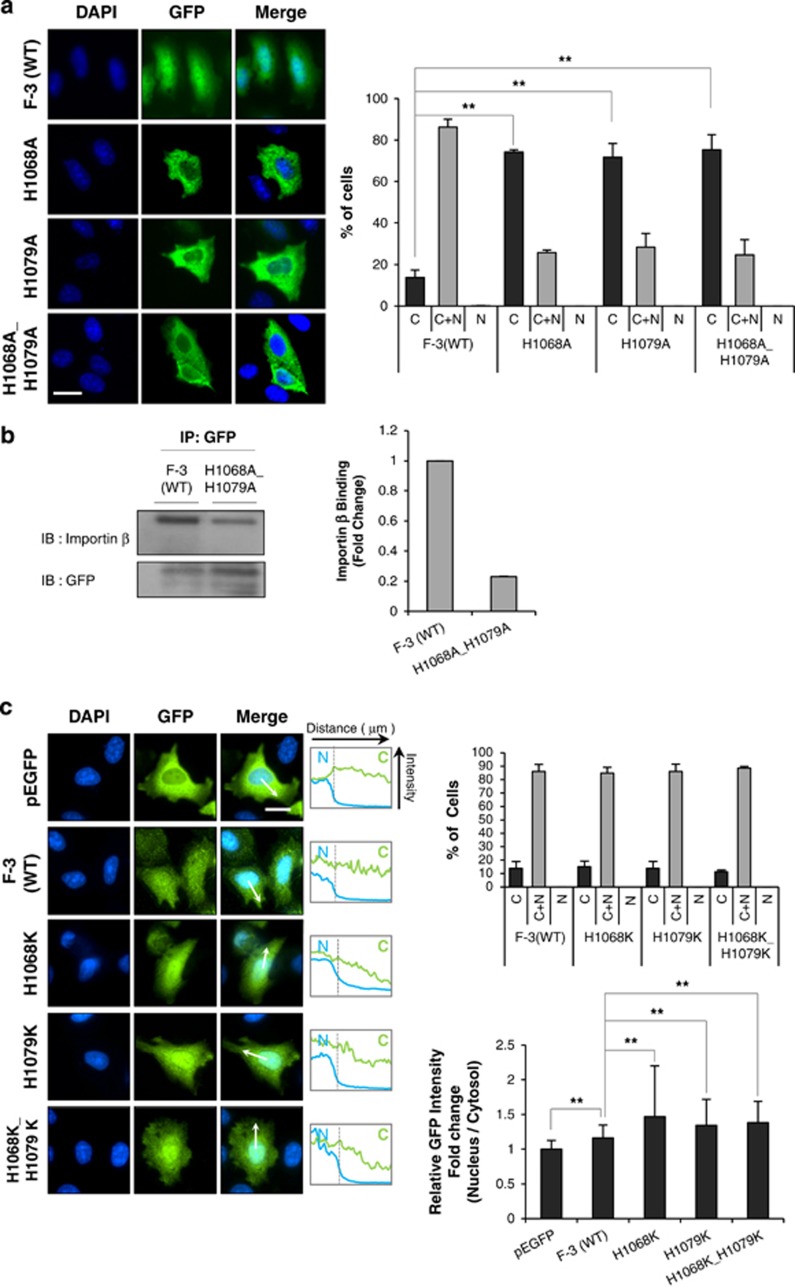
Figure 3.
Two histidine residues are crucial for the nuclear translocation of c-Met fragment. (a) HeLa cells were transfected with wild-type F-3, indicated histidine-to-alanine mutant constructs or pEGFP as a vector control. Immunocytochemistry by using anti-GFP antibody was performed as in Figure 2. Representative images were taken from fluorescence microscopy at × 40 objective. Bar graph on right panel shows the percentage of subcellular distribution of cells (n=120–225). ***P<0.001 by Student's t-test. (b) Cell lysates from HeLa cells transfected with F-3(WT) or H1068A_H1079A mutant were subjected to immunoprecipitation by using anti-GFP antibody, which was followed by immunoblotting with importin β antibody. Bar graph in right shows the relative binding tendency of importin β with cargo protein compared to F-3(WT). (c) HeLa cells were transfected with wild-type F-3 or indicated histidine-to-lysine mutant constructs, and subjected to immunocytochemistry by using anti-GFP antibodies. Bar graph displaying on right upper panel shows the percentage of subcellular distribution of cells (n=102–156) and on the right lower panel shows the relative nucleo-cytoplasmic ratio of green fluorescence intensity. Middle panel shows the intensity changes revealed by line scan analysis performed following the arrow. Error bars; standard deviations of three independent experiments. *P<0.05, **P<0.01 by Student's t-test, n=25.

