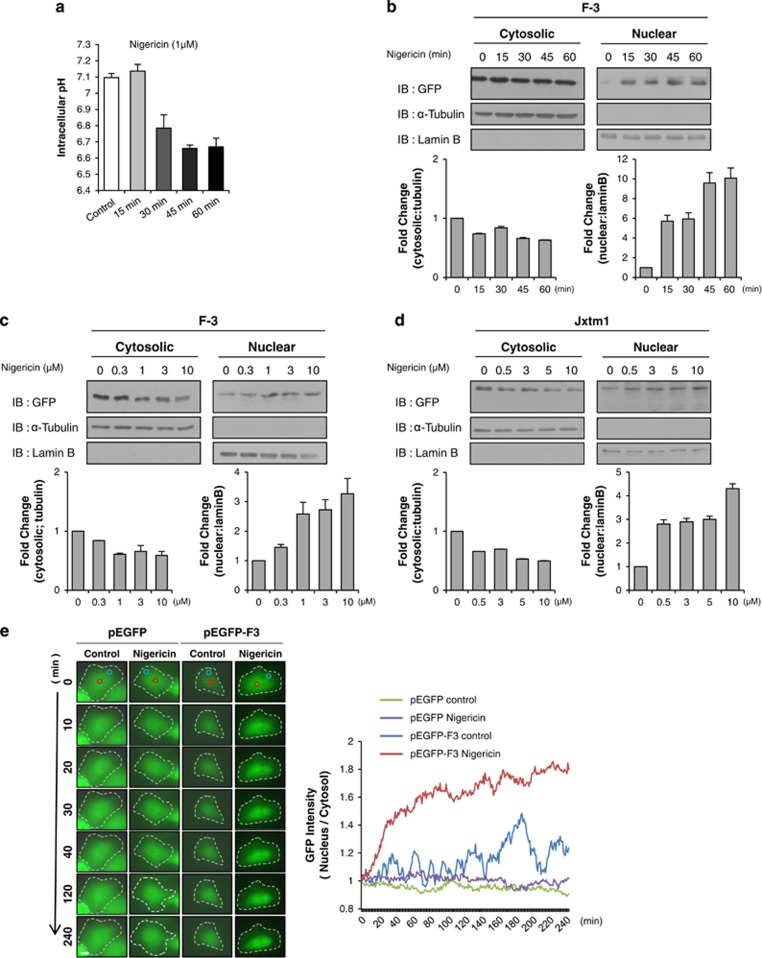
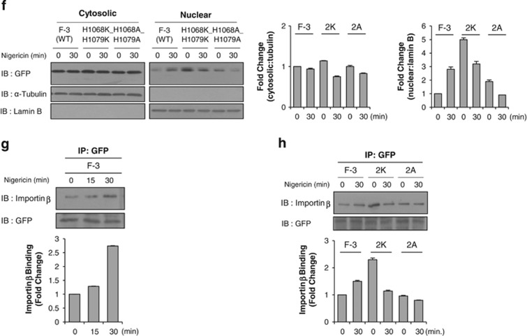
Figure 4.
Nuclear accumulation of c-Met fragment is cytosolic pH-dependent. (a) Bar graph shows the intracellular pH change measured by BCECF dye as described in Materials and methods with 1 μM nigericin treatment in a time-dependent manner. Representative result from three independent experiments. pH value of each experiment is the average of data from more than 15 cells on a cover-slip. (b) HeLa cells transiently transfected with F-3 construct were treated with or without 1 μM nigericin at indicated time intervals. Western blot analysis was performed using anti-GFP antibody after cellular fractionation. Tubulin and lamin B were used as cytoplasmic and nuclear markers respectively. Bar graphs below each panel show their respective subcellular distribution of the protein in fold change. (c, d) Western blot analysis using anti-GFP antibody was performed after subcellular fractionation of F-3 (in c) or Jxtm1 (in d)-transfected HeLa cells treated with nigericin at indicated concentration for 30 min. Bar graphs below show the subcellular distribution of the protein in fold change. (e) Representative time-lapse microscopy images of either pEGFP or F-3 transfected HeLa cells treated with 1 μM nigericin at indicated time intervals. The graph displaying on right shows nucleo-cytoplasmic ratio of GFP intensity over time. Red circle; nuclear area, white circle; cytoplasmic area. Margin of the cells are indicated by white broken line, n=10. (f) Western blot analysis using indicated antibodies were performed after subcellular fractionation of F-3 (WT), histidine-to-lysine (2K), or histidine-to-alanine (2A) mutants-transfected HeLa cells treated with or without 1 μM nigericin for 30 min. Bar graphs on right panel show the subcellular distribution of the protein in fold change. (g) Importin β binding assay (as described in Figure 3b) was performed in F-3-transfected HeLa cells after the treatment with nigericin at indicated time points. Bar graph below shows the amount of cargo-binding importin β in fold change. (h) Immunoprecipitation was performed with anti-GFP antibody with the cell lysate of F-3 (WT), histidine-to-lysine (2K) or histidine-to-alanine (2A) mutants-transfected HeLa cells treated with nigericin for 30 min followed by immunoblotting with importin β antibody. Bar graph below shows amount of cargo-binding importin β in fold change.


