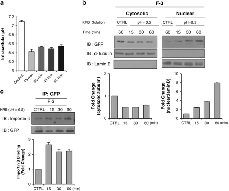
Figure 5.
Low pH KRB buffer solution enhances the nuclear accumulation of c-Met fragment. (a) Bar graph shows the intracellular pH change measured by BCECF dye as described in Materials and Methods after the treatment with low pH-KRB (pH 6.5) at indicated time intervals. Representative result from three independent experiments. pH value of each experiment is the average of data from more than 15 cells on a coverslip. (b) HeLa cells transiently transfected with F-3 construct were treated with KRB (pH=6.5) solution for indicated durations. Immunoblotting of the cell lysates after subcellular fractionation was performed using anti-GFP antibody. Bar graphs below show the changes in fold of densitometric intensity compared to control with neutral pH KRB solution (CTRL). (c) Importin binding assay was performed in F-3 – transfected HeLa cells treated with low pH buffer for indicated durations. For this, immunoprecipitation was carried out in the same way as mentioned above. Densitometric bar graph shows the binding amount of importin β (in fold change) with cargo.

