
Figure 5.
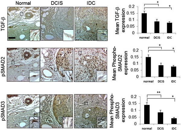
Expression patterns of CXCL1 and TGF-β signaling proteins in breast cancer stroma. Adjacent sections of normal breast (n=54) and invasive breast carcinoma (n=57) on TMAs were subject to immunohistochemistry staining for TGF-β, phosphorylated SMAD2 and phosphorylated SMAD3 proteins. Magnified insets show representative staining in fibroblastic cells. Expression was quantified by Image J, arbitrary units. Scale bar =50 microns. Statistical analysis was performed using Kruskall-Wallis tests, followed by Dunn’s post-hoc comparison. Statistical significance was determined by p <0.05. *p ≤0.001, **p ≤0.05. Values are expressed as Mean ± SEM.