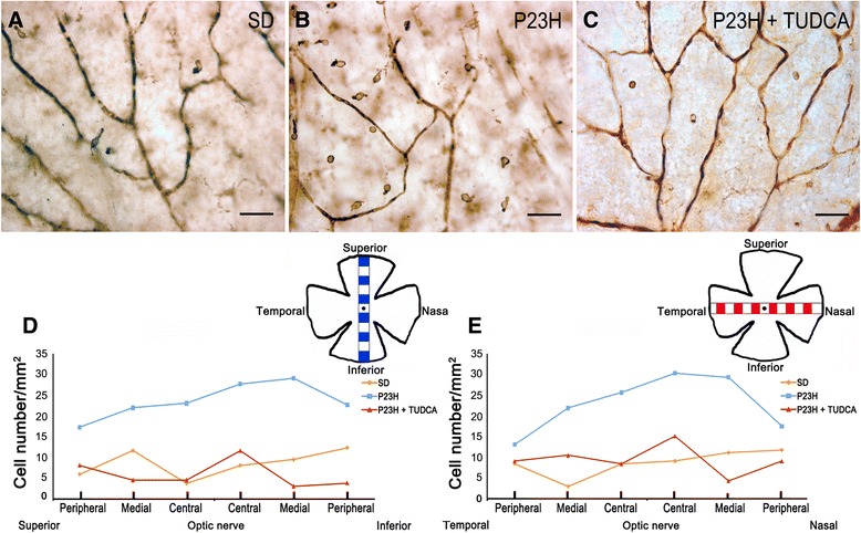
Figure 11.

Number and distribution of macrophages in the ganglion cell layer (GCL). (A- C) Representative images of whole-mount rat retinas from a SD (A), untreated P23H (B ) and tauroursodeoxycholic acid (TUDCA)-treated P23H rat (C) labeled with immunoperoxidase. (D, E) Density of macrophages, expressed as number of cells per mm2, in each of 12 representative regions in each retina: 6 equidistantly arranged on the superior-inferior axis of the retina (D) and 6 disposed on the temporal-nasal axis (E). The scheme in the margin of each panel represents the position in the retina of each representative region analyzed. Scale bar: 40 μm.
