. 2014 Oct 8;3:e03842. doi: 10.7554/eLife.03842

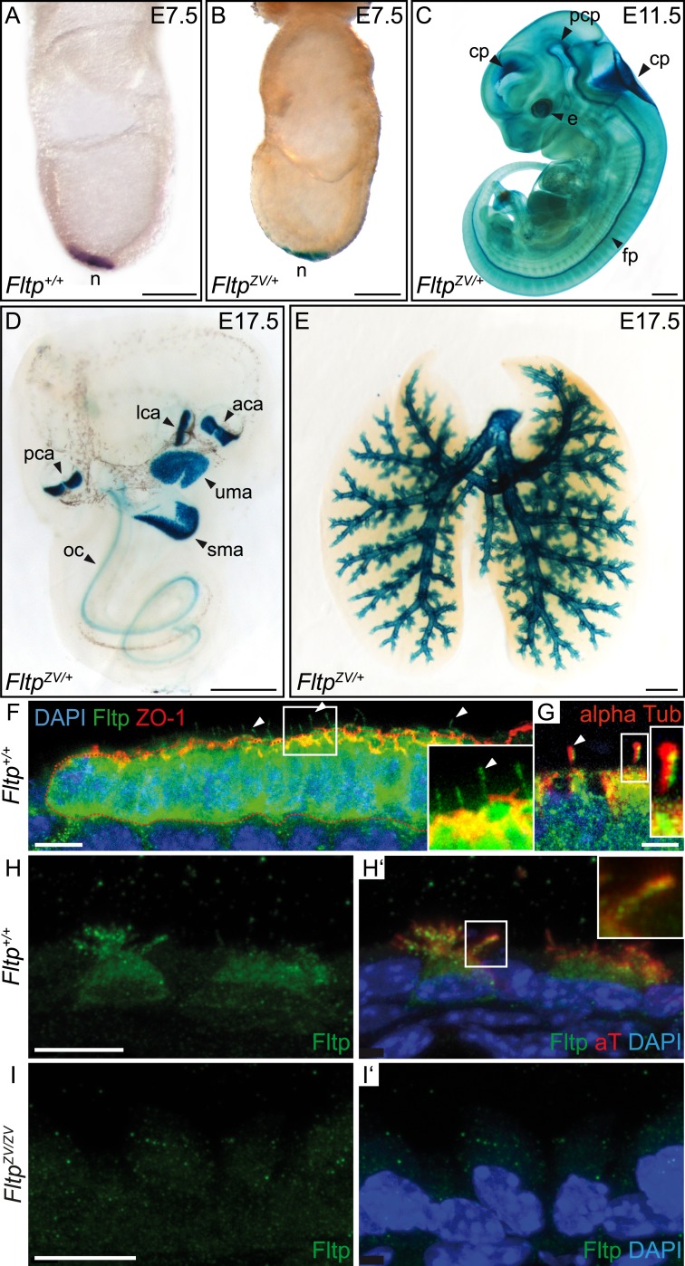
Figure 2. Fltp reporter and protein is detectable in mono- and multiciliated tissues.

(A and B) mRNA (Fltp in situ hybridization) (A) and lacZ reporter expression (B) are restricted to the node (n) at E7.5. (CE)Whole-mount lacZ stained and benzyl alcohol/benzyl benzoate (BABB) cleared FltpZV/+ embryo and organs. (C) E11.5 embryo reveals reporter expression in the choroid plexi (cp), prechordal plate (pcp), eye (e), and floor plate (fp). (D) E17.5 IE shows reporter expression in posterior crista ampullaris (pca), lateral crista ampullaris (lca), anterior crista ampullaris (aca), utricular macula (uma), and saccular macula (sma) of the vestibular part as well as in the organ of Corti (oc). (E) E17.5 lung shows lacZ reporter activity in multiciliated lung epithelial cells. (F and G) Whole-mount antibody stained embryo (node, E7.75) analyzed by LSM reveals Fltp protein in vesicular fashion in the cytoplasm and along primary cilia (white arrow heads). (HI′) Immunohistochemistry on cryosections combined with LSM analysis reveals Fltp at the apical plasma membrane and at cilia in multiciliated lung epithelial cells of Fltp+/+adult animals (H and H′). No Fltp immunoreactivity is detected in FltpZV/ZV lungs (I and I′). ZO-1 marks apical TJs, α-Tubulin (alpha Tub), and acetylated-Tubulin (aT), the tubulin network and cilia; nuclei are marked by DAPI and Fltp by Fltp1 (F and G) and Fltp116-1 (HI′). Scale bars; 100 µm (A and B), 500 µm (C, D, E), 10 µm (F), 4 µm (G), and 10 µm (HI′).

DOI: http://dx.doi.org/10.7554/eLife.03842.007

Figure 2.

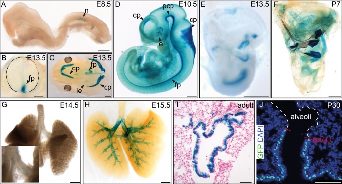
Figure 2—figure supplement 1. Additional Fltp reporter expression in mono- and multiciliated tissues.

Figure 2—figure supplement 1.

(AH) Whole-mount lacZ stained and benzyl alcohol/benzyl benzoate (BABB) cleared (D, F, H) FltpZV/+ embryos and organs. β-gal activity is restricted to the node (n) (A). (B) Cross-section of the neural tube (dotted line) reveals reporter expression in the floor plate (fp). (C) Reporter expression can be detected in the four choroid plexi (cp), the fp, and the inner ear (ie). (D) lacZ expression analysis reveals β-gal activity in the developing cp of the first, second, third, and fourth ventricle, in the prechordal plate (pcp), in the developing eye (e), and in the fp. (E and F) LacZ reporter is expressed in the 6 sensory regions of the IE. Onset of reporter expression is at E12.5. (G and H) LacZ staining of whole-mount lungs shows onset of reporter expression in the main stem bronchi at E14.5 (G). Later, expression is restricted to the lung epithelium (H). (I) Histological section through a lacZ stained adult lung showing β-gal activity in the lung epithelium. (J) Laser scanning microscopy of lung cryosections reveals Fltp reporter expression in epithelial lung cells. Expression directly stops at the broncho-alveolar-duct junction (BADJ). The alveoli are absent of Fltp reporter staining. Fltp reporter positive cells are marked by GFP and nuclei by DAPI. Scale bars; 200 µm (A), 250 µm (B), 500 µm (CH), 150 µm (I), 75 µm (J).