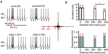
Figure 5. A model neuron based on the homogeneous properties of the entire dendrite shows substantial contribution of a distal dendrite to pacemaker activities.

A, current injection to a various part of a model DA neuron affected spontaneous firing activities: firing enhancement during the current injection and post-stimulatory subsequent pause of firing. Schematic shape of a model neuron and each stimulation site are presented in the centre. The red spots of the model neuron showed current injected locations. A 40 pA current was injected for 500 ms. B, firing changes were classified into excitation during the stimulation and post-stimulatory inhibitions. Bar graphs represent the firing frequencies of excitation (upper) or inhibition (bottom) responses (*P = 0.02).
