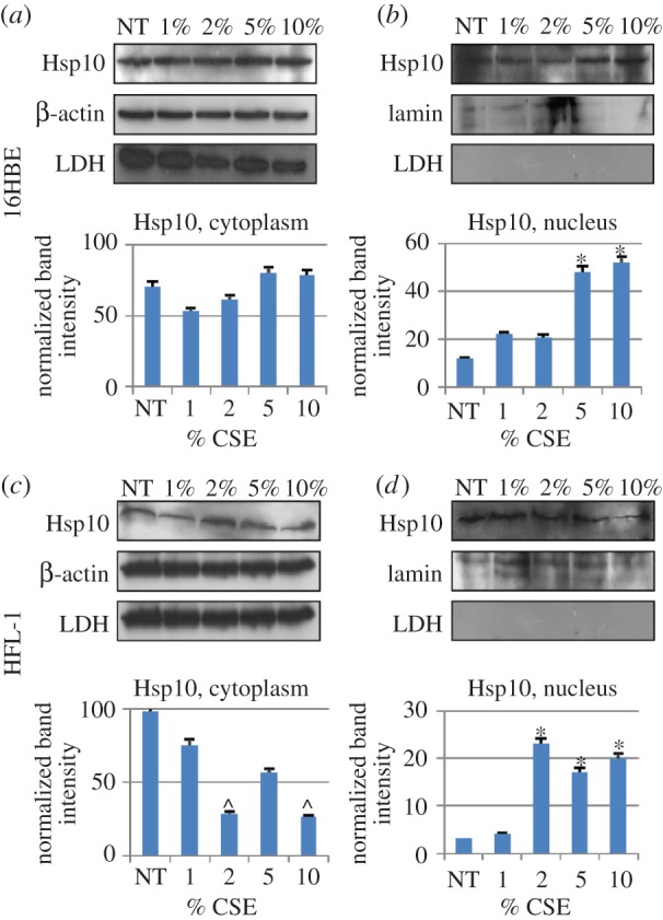
Figure 6.

Identification of Hsp10 in subcellular fractions by western blotting. Hsp10 was detected in the cytoplasmic fraction (a,c) of 16HBE (a) and HFL-1 (c) cells treated with various concentrations of CSE (from 1 to 10%, as indicated). Likewise, Hsp10 was detected in the nuclear fraction of both cells lines (b,d) and its levels were significantly higher after treatment with 2% or higher concentrations of CSE by comparison with NT cells in HFL-1 (p < 0.05), and 5% or higher in 16HBE (p < 0.05), indicating that this treatment induced an increase in Hsp10 in the nucleus. A significant (p < 0.05) decrease in Hsp10 was observed in the cytoplasm of HFL-1 cells after treatment with 2 and 10% CSE. β-Actin and lamin were used as cytoplasmic and nuclear internal controls, respectively. Possible contamination of nuclear preparations with cytoplasm was monitored by testing for lactate dehydrogenase (LDH), but no contamination was detected.
