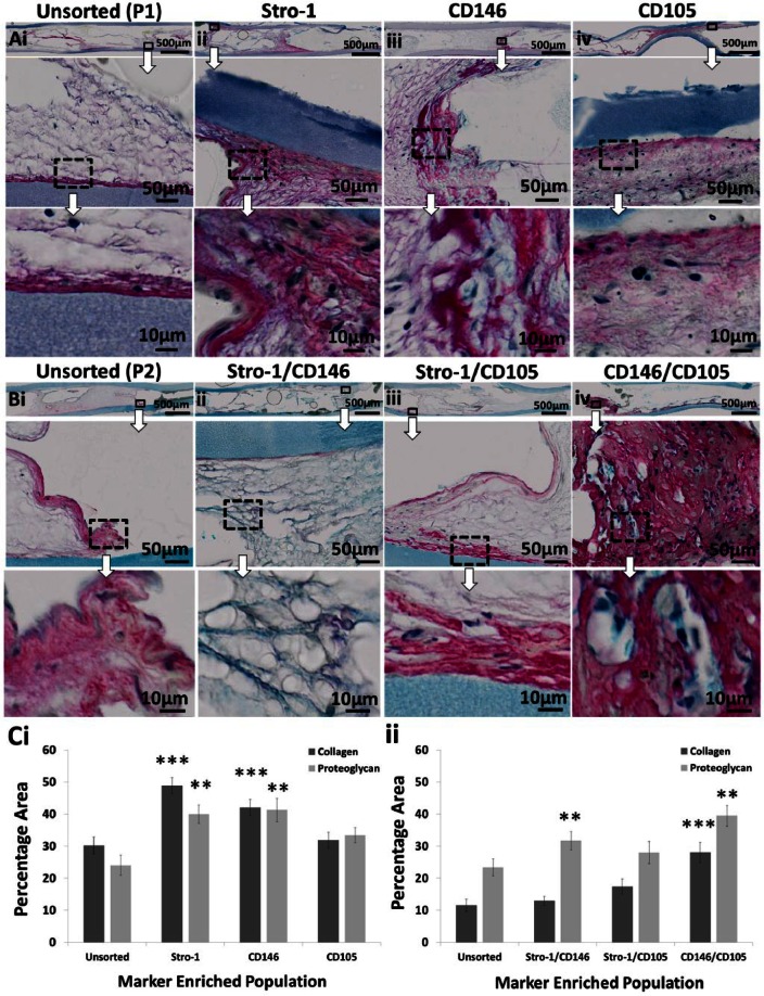
Figure 7.
Histological assessment of single- and dual-labelled population viability/function in vivo. (Ai and Bi) Unsorted, (Aii to Aiv) single- and (Bii to Biv) dual-labelled populations were expanded in vitro and seeded as a cell suspension into diffusion chambers at 5 × 10⁵ cells per chamber without osteoinduction. Chambers were implanted subcutaneously within MF1 nu/nu mice and incubated for 28 days. Following harvest, chambers were sectioned and stained with Alcian Blue/Sirius Red. Collagen and proteoglycan were identified and quantified by image analysis and represented as a percentage of the total sample (C). Overview images of the entire implant are shown on top (scale bar 500 µm). Large images underneath show enlarged regions (scale bar 50 µm). Highly magnified images are shown at the bottom (scale bar 10 µm). Error bars are SD (**p ≤ 0.01, ***p ≤ 0.001).
SD: standard deviation.

