
Figure 2.
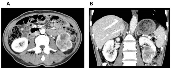
Abdominal enhanced computed tomography (CT) after axitinib treatment. (A, axial section; B, coronal section). Abdominal CT shows a hypervascular tumor (size, 4.5 × 2.5 cm) in the lower pole of the left kidney. The tumor diameter shrank by 56%, compared to that before axitinib treatment.